
Figure 3. Transient appearance of type IIA fibres in denervated and 20 Hz stimulated EDL muscles.
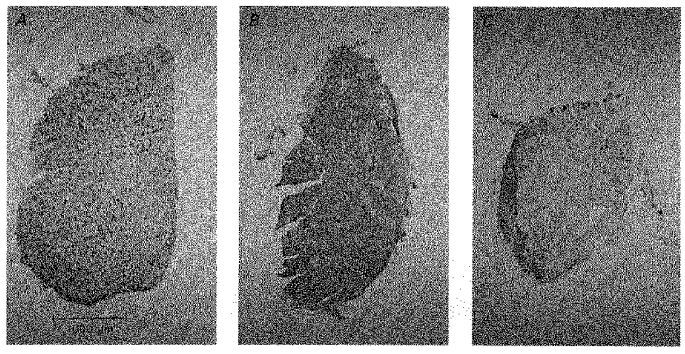
Cross-sections of EDL muscles after 10 (A), 43 (B), and 124 (C) days of denervation plus direct stimulation. The sections were stained with the BA-71 antibody against type IIA MHC.