
Figure 5. Effect of ATP in the presence and absence of extracellular Ca2+.
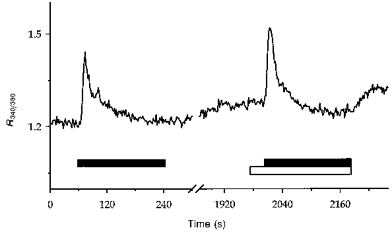
Experimental procedures were as described for Fig. 1. A PCT was exposed to 100 μmol l−1 ATP for the periods indicated by the horizontal filled bars successively in the presence (1.8 mmol l−1 CaCl2) and absence (100 μmol l−1 EGTA, open bar) of external Ca2+. The break in the trace represents a 25 min period of superfusion with Ringer solution.