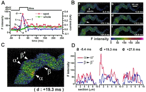
Figure 4. Ca2+ image and membrane current during depolarization to -20 mV in a single myocyte of guinea-pig urinary bladder.

A, membrane potential of a cell stepped from -60 to -20 mV. Ca2+ current and subsequent transient outward current were observed (blue dots). Green circles indicate the averaged F intensity over the whole area of the cell. Red triangles indicate F intensity in a hot spot (a circle of 1.2 μm in diameter) in a subplasmalemma area at the position indicated by an arrow in Bc. The images were obtained at 6 different times as indicated (a-f) and are shown correspondingly in B and D. Transient outward current was activated with the same time course as the rise of [Ca2+] in the hot spot. Moreover, the increase in hot spot F intensity was also transient and decreased just after repolarization to a level slightly higher than that at rest. B, Ca2+ images obtained at the times shown in A. The recording pipette was placed at the centre of the cell, indicated by an arrow in a.C, the image shown in Bd (+19.3 ms) is enlarged. D, profiles of F intensity along two cross-sections indicated by the two sets of small triangles in C; α-α′ and β-β′ are plotted against the cell section (red and blue lines, respectively). Profiles in a, d and e were obtained from corresponding images in B. Zero corresponds to the position of the cell membrane. The line α-α′ was placed just on the centre of the hot spot and the line β-β′ was placed in a quiescent area. The peak of intensity in Dd indicates that the centre of the Ca2+ hot spot is located about 0.7 μm from the edge of the cell. In contrast, the intensity profile along line β-β′ showed only low-level fluctuations in [Ca2+]. In the presence of 0.1 mm Cd2+, both hot spots and IK,Ca were not observed on depolarization to -20 mV (not shown). Similar transient outward current and Ca2+ hot spots on depolarization to -20 mV were observed in all cells examined, 5 vas deferens and 3 urinary bladder myocytes.
