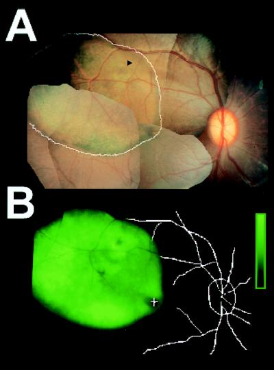
Figure 1.
In vivo imaging shows fluorescence localized to the site of subretinal rAAV.CMV.EGFP injection at 16 weeks. Montage of color photographs (A) and fluorescence intensity (B) in eye 1 of animal 94B-109 (Table 1). Extent of fluorescence from B is overlaid (white trace) on A. Arrowhead, injection site; Fundus landmarks (+, fovea) from A are overlaid on B. Fluorescence intensity is mapped to increasing intensities of green color; scale bar represents 2.4 log units above background.

