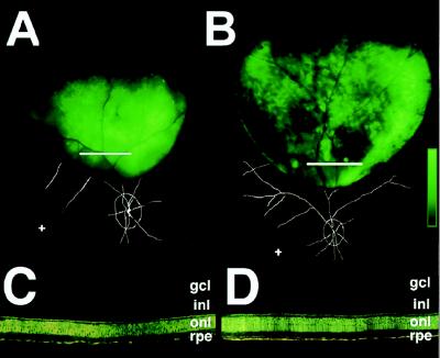
Figure 3.
Subretinal rAAV.CMV.EGFP administration to the eye contralateral to that injected with rAAV.CMV.EGFP 7 months earlier results in high levels of EGFP protein. (A and B) Fundus fluorescence 7 weeks [95C-273 eye 2 (A)] and 16 weeks [94B-051 eye 2 (B)] after readministration. Scale bar represents 2.1 log units (A) or 2.4 log units (B) of fluorescence above background. Fundus landmarks are overlaid (+, fovea). White lines in A and B indicate the regions shown histologically in C and D, respectively. gcl, ganglion cell layer; inl, inner nuclear layer; onl, outer nuclear layer; rpe, retinal pigment epithelium.

