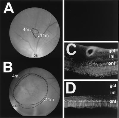
Figure 4.
EGFP fluorescence is stable over time. Shown is the extent of in vivo fluorescence at 4 and 11 months (4 m and 11 m) postinjection in 95C-273 eye 1 (A) and 94B-051 eye 1 (B). White lines in A and B indicate the regions shown histologically in C and D, respectively. ON, optic nerve. The section shown in C was thicker than that in D (30 vs. 18 μm) to enhance detection of the weaker EGFP-labeled photoreceptors in 95C-273 eye 1. Use of the thicker section results in increased retinal autofluorescence. gcl, ganglion cell layer; inl, inner nuclear layer; onl, outer nuclear layer.

