
Figure 6. Na+-HCO3− cotransport current in isolation.
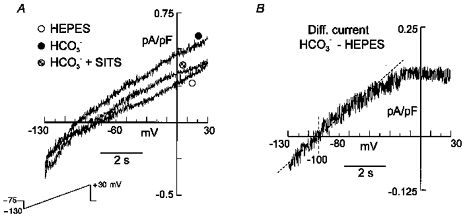
Standard whole-cell configuration. A, steady-state currents, evoked by the same voltage protocol used in Fig. 3, recorded from a rat cardiac myocyte exposed successively to external Hepes, HCO3−, and HCO3− in the presence of 0.1 mm SITS. External and internal K+, Cl− and Ca2+ were replaced with Cs+, methanesulphonate and Mg2+, respectively. TTX, nifedipine and TEA were added to the extracellular solution. TEA was also included in the pipette solution. B, HCO3−-sensitive difference current (HCO3− - Hepes) with a reversal potential of −101 mV.