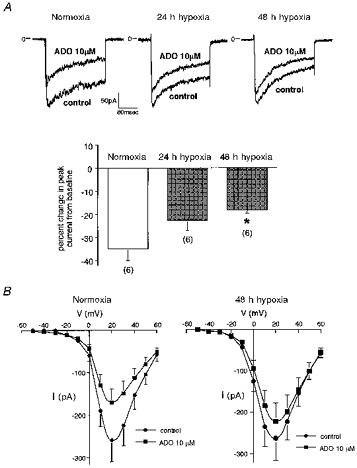
Figure 1. Attenuation of adenosine-induced inhibition of voltage-dependent Ca2+ current by long-term hypoxia in PC12 cells.

A (upper panel): traces showing the effect of adenosine (ADO) on ICa. ICa was measured every 30 s by 160 ms test pulses from a Vh of −80 to +20 mV. Peak current amplitude was measured for evaluation. The charge carrier was 20 mm Ba2+. In normoxic controls, adenosine (10 μm) elicited a decrease in the amplitude of ICa (left). This effect of adenosine was smaller when the cells had been pretreated with hypoxia (10 % O2) for 24 h (middle) or 48 h (right). A (lower panel): the effect of adenosine on ICa was significantly attenuated when the cells had been exposed to 10 % hypoxia for 48 h (*P < 0.05). The response to adenosine was evaluated as the percentage inhibition from baseline inward current. The numbers in parentheses indicate the number of cells examined. Means + s.e.m. are shown. B, effect of adenosine (ADO) on the peak current-voltage relationship of ICa. Means + s.e.m. are shown (n = 5 for each group). ICa was measured with 100 ms long test pulses from a Vh of −80 mV to test potentials ranging from −50 to +60 mV (10 mV increments). Peak currents were measured before and after steady-state inhibition by the application of adenosine (10 μm). Adenosine decreased ICa at the voltage range examined.
