
Figure 7. Three examples of premature bifurcation of the main apical dendrite in the irradiated cortex.
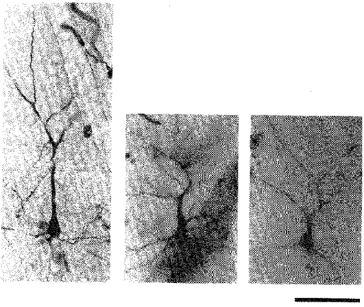
Photomicrographs of HRP-stained neurones in coronal sections. In each panel the soma is in the lower half and the apical dendrite extends upwards, and soon divides. Scale bar denotes 100 μm.