
Figure 1. Fractional inhibition of labelled D-Phe-L-Gln (0.42 μm) influx.
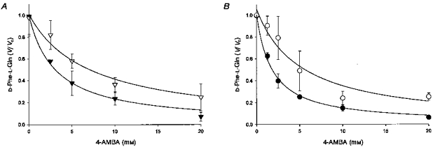
A, fractional inhibition (V/V0) of labelled D-Phe-L-Gln (0.42 μm) influx into PepT1-expressing Xenopus laevis oocytes by 4-AMBA (0–20 mm) when pHo = 5.5. Continuous lines represent the best fit of data to Michaelis-Menten kinetics for a single system in the absence (▾) and presence (▿) of unlabelled D-Phe-l-Gln (1.0 mm). Calculated inhibition constants (Ki) are 3.1 ± 0.4 and 6.9 ± 1.2 mm, respectively. Data has been corrected for non-mediated transport by subtracting D-Phe-L-Gln flux seen in H2O-injected control oocytes. Labelled D-Phe-L-Gln flux in the absence of 4-AMBA has been normalized to 1. Absolute fluxes in the absence and presence of unlabelled D-Phe-L-Gln are 194 ± 36 and 118 ± 18 fmol oocyte−1 h−1, respectively. Data points are means ± s.e.m. (n = 5). B, fractional inhibition of labelled D-Phe-L-Gln (0.34 μm) influx into rat renal BBMV by 4-AMBA (0–20 mm) under equilibrium exchange conditions: pHo = 5.5, pHi = 7.4, 4-AMBAo = 4-AMBAi and membrane potential clamped inside negative. Continuous lines represent the best fit of data to Michaelis-Menten kinetics for a single system in the absence (•) and presence (^) of unlabelled D-Phe-l-Gln (0.21 mm). Calculated inhibition constants (Ki) are 1.8 ± 0.1 and 5.1 ± 1.3 mm, respectively. Data has been corrected for non-mediated transport by subtracting labelled D-Phe-L-Gln flux seen in the presence of a saturating concentration of unlabelled peptide (10 mm). Labelled D-Phe-L-Gln flux in the absence of 4-AMBA (Vo) has been normalized to 1. Absolute fluxes in the absence and presence of unlabelled D-Phe-L-Gln are 854 ± 227 and 464 ± 208 fmol (mg protein)−1 s−1, respectively. Data points are means ± s.e.m. (n = 3–6).