
Figure 6. Ryanodine induced subplasmalemmal Ca2+ increase in bovine coronary endothelial cells monitored by FFP-18.
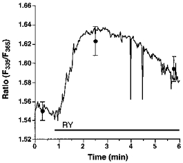
Endothelial cells freshly isolated from bovine circumflex coronary artery were loaded with FFP-18 using laser-generated stress waves to monitor changes in [Ca2+]sp. After 10 min equilibration time at room temperature, cells were washed twice, resuspended in Ca2+-free HBS and placed into a stirred cuvette. Changes in [Ca2+]sp were monitored at 335 and 365 nm excitation and 500 nm emission. Each point represents the mean ±s.e.m. (n= 14).