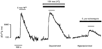
Figure 8. Membrane potential modulates Ni2+-induced cytosolic Ca2+ transients.

Representative traces showing the effect of extracellular [Ni2+] (5 mM; black lines) on the cytosolic [Ca2+] of cultured TM3 cells, in various protocols whereby the membrane potential was altered by using the K+ ionophore, valinomycin (5 μM; filled bars), in the presence of either 5 mM (hyperpolarized) or 100 mM [K+]e (open bar) (depolarized).
