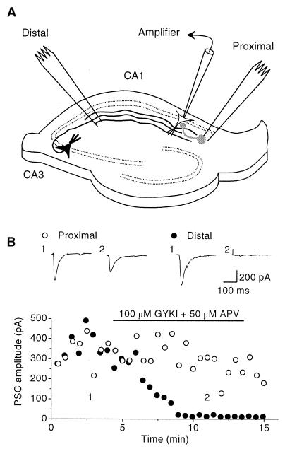
Figure 1.
Experimental design. (A) Schematic illustration of the hippocampal slice showing the positioning of the recording pipette and stimulating electrodes. Schaffer collateral axons are shown in black, and a local inhibitory interneuron is shown in gray. (B) Effect of blocking AMPA and NMDA receptors with GYKI52466 and APV, respectively. The response to the distal stimulus (●) was completely blocked. The proximal stimulus (○), however, continued to elicit a monosynaptic response. Insets show averages of five successive traces obtained at the times indicated.

