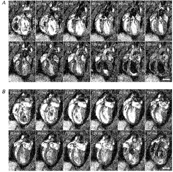
Figure 2. Longitudinal cardiac sections.

A series of typical longitudinal sections taken parallel to the principal cardiac axis at one spatial slice obtained from a normal male WKY weighing 300 g and aged 12 weeks (A) and from a male SHR weighing 319 g and aged 12 weeks (B). The intrinsic heart rates of these rats were 331 ± 3 and 287 ± 7 beats min−1, respectively. Each image is the average of two signals obtained at corresponding points in the cardiac cycle following the R wave. The time indicated in the upper left-hand corner of each panel is the delay after the trigger, taken from the R wave, at which each signal was acquired. The left and right ventricles (LV and RV), atria (LA and RA) and the aorta (AO) are labelled. The nominal in-plane resolutions (pixel sizes) were 350 and 390 μm for the WKY and SHR, respectively, and slice thicknesses were 1.12 mm for both animals. For display, the images have been zero-padded from a matrix of 128 to 256 pixels square. The effective repeat time was approximately 400 ms. The scale bars indicate 5 mm.
