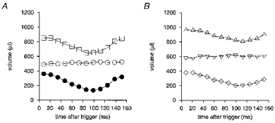
Figure 6. Time series of left ventricular volume.

Endo-, epi- and myocardial LV volumes for one typical WKY rat (A;□, epicardial; ○, myocardial; •, endocardial) and one typical SHR (B;▵, epicardial; ▿, myocardial; ⋄, endocardial) plotted against time after the R wave trigger, measured from transverse MR sections. Heart rates were 293 ± 3 beats min−1 for the SHR and 304 ± 7 beats min−1 for the WKY. The SHR and WKY rats used for measuring these volumes had very similar body weights (241 and 239 g, respectively). The LV myocardial volume was greater in the SHR and its ejection fraction was reduced, as indicated by the raised end-systolic volume compared with the WKY. This is consistent with the overall parameters for each group of animals specified in Table 1.
