
Figure 5. Low K+-induced membrane potential changes in Na+-free and Cl−-free solutions and in the presence of 10 mm[EGTA]i.
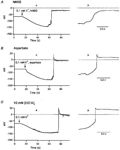
A, effect of 0.1 mm[K+]o in the absence of extracellular Na+; at the arrow, superfusion was switched from normal Tyrode solution to low K+, Na+-free (NMDG) solution. B, effect of 0.1 mm[K+]o in the absence of extracellular Cl−; at the arrow, superfusion was switched from normal Tyrode solution to low K+, Cl−-free (aspartate) solution. C, 0.1 mm[K+]o solution was introduced at the arrow, and membrane potentials were recorded with a pipette containing 10 mm EGTA. Introduction of 0.1 mm[K+]o caused hyperpolarization, irregular, transient depolarizations and sustained action potentials. Panels labelled a depict changes in Vrest values and an action potential on a slow time scale, and panels labelled b depict the rising phases of the action potential on a faster time scale.