
Figure 1. AP waveform to illustrate the variables measured.
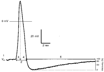
An intracellularly recorded somatic AP of an Aα/β-fibre neurone evoked by electrical stimulation of the dorsal root showing the electrophysiological parameters measured. These were membrane potential (Vm) (1), AP duration at base (APdB) (2), AP rise time (RT) (3), AP fall time (FT) (4), and AHP duration to 80 % recovery (AHP80) (5).