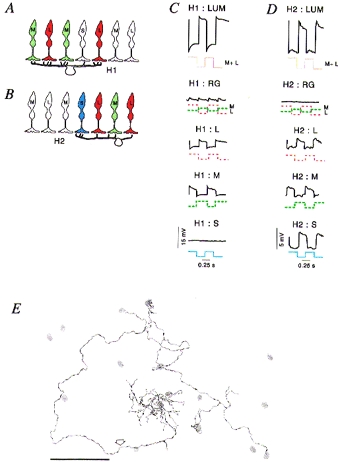
Figure 2. Connectivity and responses of horizontal cells.

A and B, schematic views of vertical sections through primate retina showing the connection patterns of the dendrites of H1 and H2 horizontal cells. The H1 cells (A) make only very sparse contact with S cones within their dendritic field, but the H2 cells (B) contact all cone types, and make stronger connections with S cones. C and D, response of H1 and H2 cells in macaque retina, respectively. Each graph shows intracellular recordings of a single H1 or H2 cell to temporal square wave modulation of L, m or S cones types. Only the H2 cell shows measurable input from S cones. E, camera lucida drawing of an H2 cell in marmoset retina, showing the position of S cones (grey areas) within the dendritic and axonal field of the cell. The dendrites of the H2 cell make substantial contact with two S cones within the field. The axon (arrow) also makes contact with S cones which lie along its path. Panels C and D are modified from Dacey et al. (1996); panel E is modified from Chan & Grünert (1998). Scale bar in E, 50 μm.
