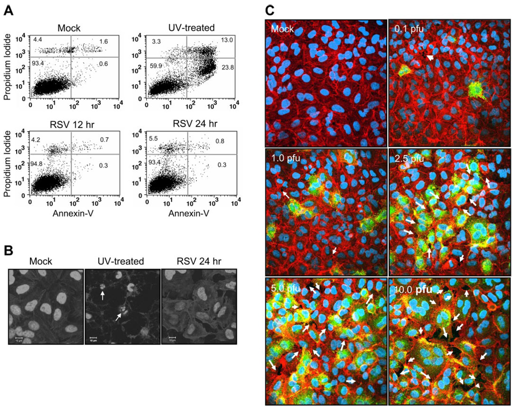
Fig. 2.
RSV infection does not cause apoptosis but causes paracellular gaps. A549 cells were treated with vehicle alone (Mock), treated with UV (30 min) to induce apoptosis, or infected with RSV at multiplicity of infection (MOI) of 2.5 pfu/cell. Apoptosis levels were then determined after further incubation by annexin V staining and flow cytometry (A) or with 4,6-diamidino-2-phenylindole (DAPI) nuclear staining (B) and confocal microscopy (n = 3). Scale bar represents 10 µm. C: to test for the presence of paracellular gaps, confluent monolayers of A549 cells were infected with RSV at concentrations indicated in the figure. The cells were then stained with Texas red-phalloidin for actin and DAPI for nuclear staining. RSV-infected cells were visualized by using FITC-labeled goat anti-RSV antibody. Arrows indicate paracellular gaps (n = 2).

