Figure 3.

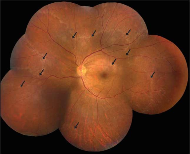
Montage of retinal photographs of the left eye of patient 2 during initial admission demonstrating a 360° subretinal whitish-gray ring along the retinal midperiphery (arrows).

Montage of retinal photographs of the left eye of patient 2 during initial admission demonstrating a 360° subretinal whitish-gray ring along the retinal midperiphery (arrows).