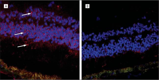
Figure 4.

A, Using the same technique as for patient 1, indirect immunohistochemistry with confocal microscopy–enhanced imaging was performed in patient 2 and showed positive staining (arrows) with serum localized to the inner and outer nuclear layers, with faint staining along the inner segments of the photoreceptors. B, There was no definite reaction using serum from a healthy control as the primary antibody and goat antihuman Cy3 red color as the secondary antibody on a normal unfixed, frozen human retina (blue, 4′,6-diamidino-2-phenylindole dihydrochloride staining for nuclei; green, autofluorescent staining for retinal pigment epithelium lipofuscin; original magnification × 250).
