
Figure 2.
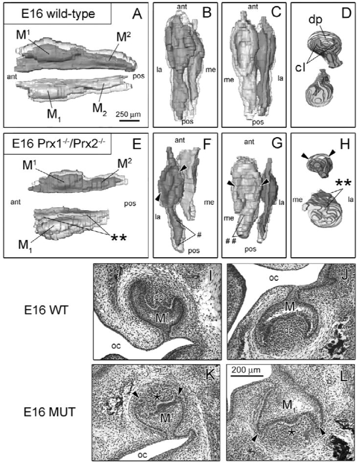
Prx1-/-/Prx2-/- molars show patterning defects at E16. (A color version of this Fig. is included in the APPENDIX.) WT (n = 3) and MUT (n = 3) 3D reconstructions (A-H) are presented from various viewpoints: lateral (A,E), superior (B,F), inferior (C,G), and frontal (D,H). Histological images of frontal sections are displayed for WT mice (I,J) and MUT (K,L). Morphological differences between WT and MUT include hypoplastic cervical loops (cl) (arrowheads), and dental papilla (dp) in the MUT-M1 and MUT-M1 are hypoplastic. In addition, MUT-M1 epithelium is more malformed and larger than the corresponding M1. WT-M1 is still inclined medially, while MUT-M1 lacks this positional displacement. WT maxillary (M2) and mandibular (M2) second molars are developing posterior to bell-stage WT-M1 and WT-M1, while MUT-M2 is hypoplastic (#) and MUT-M2 is absent (##). Key: oral cavity (oc), is anterior (ant), posterior (pos), medial (me), lateral (la).