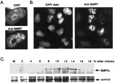
Figure 4.
Cell cycle-dependent expression of SMRTe. (A) Immunofluorescence staining of endogenous SMRTe in HeLa cells. The nucleus stained by 4′,6-diamidino-2-phenylindole (DAPI) dihydrochloride hydrate (Upper) and the SMRTe signal (Lower) is shown. Note that SMRTe is distributed as fine granules in the nucleus and is excluded from the nucleoli. (B) Immunostaining of SMRTe in an unsynchronized population of A549 cells. Strong SMRTe signals were detected in a subset of A549 cells. (C) Western blotting for SMRTe in A549 cells at different time points after release from mitosis. The 270-kDa SMRTe signal (Upper) and a nonspecific band (Lower) are shown.

