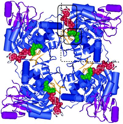
Figure 1.
Human Type II IMPDH tetramer with bound dinucleotide analogue SAD (circled, red) and substrate analogue 6-Cl-IMP (circled, green). The dinucleotide binds at the monomer–monomer interface (dotted lines). The following structures are illustrated: catalytic β-barrel domain (blue), flanking domain (magenta), active site loop (yellow) and active site flap fragments (white).

