
Figure 1. Brain–skin cross-talk upon exposure to psychological stress or environmental stress factors.
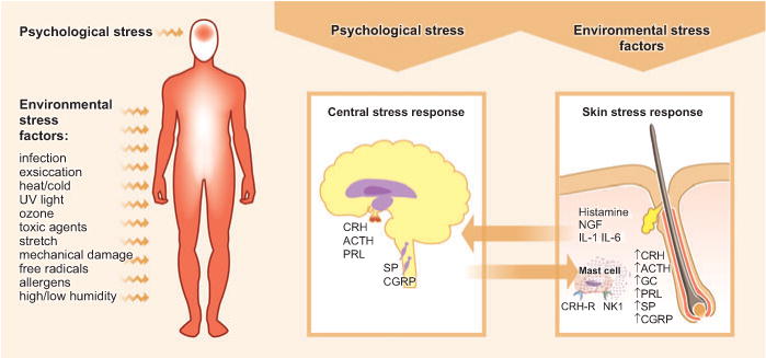
Upon perception of psychological stress, the central stress response leads to the activation of the hypothalamic–pituitary–adrenal axis, which causes the release of corticotropin-releasing hormone (CRH), ACTH, and prolactin (PRL). Further, an upregulation of substance P (SP) and calcitonin gene-related peptide (CGRP) can be observed in the dorsal root ganglia. Such stress response patterns may be translated into a skin stress response, including the local production of CRH, ACTH, and glucocorticoids (GCs), the release of inflammatory cytokines, and the sprouting of SP+ nerve fibers. In the skin response to stress, mast cells occupy a central switchboard position, as they are targets for stress-triggered factors as well as effector cells that contribute, for example, to neurogenic inflammation in the skin. Environmental factors are also capable of inducing a skin stress response, and this may be signaled to the brain, where it affects behavior and leads to an increased vulnerability to additional stress perception. NGF, nerve growth factor.