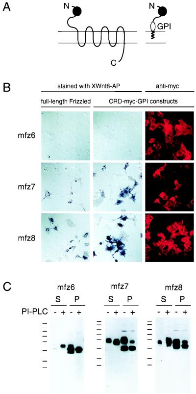
Figure 2.
Binding of XWnt8-AP to full-length Frizzled proteins and the corresponding GPI-anchored CRDs on the surface of transfected COS cells. (A) Schematic diagram showing the location of the CRD (filled ball) in a full-length Frizzled protein and in a CRD-myc-GPI construct. Horizontal lines represent the membrane, and zigzag lines represent the lipid component of GPI. N, amino terminus; C, carboxyl terminus. (B and C) XWnt8-AP binding and surface localization assays for three Frizzled proteins that show undetectable binding (mfz6), intermediate binding (mf7), and strong binding (mfz8). (B) Light microscopy of representative samples of COS cells transiently transfected with the indicated full-length Frizzled (left column) or the corresponding Frizzled CRD-myc-GPI construct (center and right columns). Live cells were stained with XWnt8-AP (left and center columns) or with anti-myc mAb and a fluorescent secondary antibody (right column). The cells remained intact during the binding reaction as determined by the failure of the anti-myc mAb to bind myc-tagged Dishevelled (a cytoplasmic protein) under these conditions; fixation and permeabilization with acetone and paraformaldehyde before incubation with the anti-myc mAb led to intense staining of myc-tagged Dishevelled (data not shown). (C) PI-PLC release of GPI-anchored CRDs from the surface of live cells. Transfected COS cells were incubated in the absence (−) or presence (+) of PI-PLC, and, after centrifugation, the released protein was recovered from the supernatant (S) and the cells were recovered from the pellet (P). Proteins were resolved by SDS/PAGE and were visualized with anti-myc mAb immunoblotting. Molecular mass standards are 194, 120, 87, 64, 52, 39, 26, and 21 kDa.

