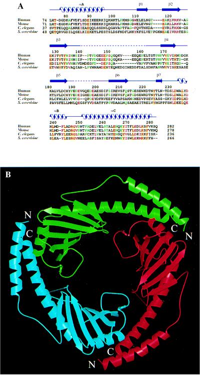
Figure 2.
Overall structure of p32. (A) Structure-based alignment of p32 sequences from human (PID g338043), mouse (PID g743485), Caenorhabditis elegans (PID g3334445) and S. cerevisiae (PID g557799). Only the sequences corresponding to the mature human protein are shown. Positions with four identical amino acids are shown in red letters over yellow background; positions with four similar amino acids are shown in plain red letters; positions with three identities are shown in green letters. Related expressed sequence tag fragments or genomic sequences can also be found in Drosophila (NID g3112239), zebrafish (NID g2446858), and Arabidopsis (PID g3334441). PID and NID are protein and nucleotide identification numbers in GenBank. (B) Ribbon representation of the p32 trimer, looking down the noncrystallographic three-fold axis. The dotted lines show disordered segments in the structure. The three monomers that make up the trimer are colored red, green and cyan, and are referred to as subunits A, B, and C, respectively. The same color codes are used in other figures.

