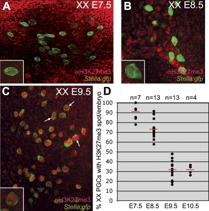
Figure 3. During PGC Migration the H3K27me3 Nuclear Accumulation Is Gradually Lost.
The total number of PGCs per embryo was analysed in XX Stella:gfp embryos after whole mount immunostaining for H3K27me3. Stella is a marker of specified PGCs.
(A–C) E7.5 (A), E8.5 (B), and E9.5 (C) confocal projections showing all PGCs present in the depicted area. Magnification of single Stella(GFP)-positive PGCs containing a prominent nuclear H3K27me3 accumulation are depicted in the lower right corner of (A–C). Note that the overall levels of H3K27me3 in the nucleus increase during development (white arrows in C).
(D) The percentage of PGCs containing a prominent H3K27me3 accumulation from E7.5 to E10.5. Red bars depict the median, n is the total number of embryos analysed.

