
Figure 1.
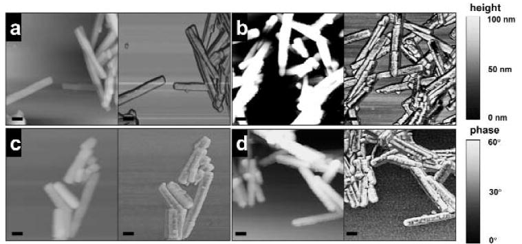
Tapping mode AFM images of enamel crystals on a mica surface, imaged in air. (a) Control (maturation stage); (b) maturation-stage enamel crystals from rats after systemic administration of 50 ppm fluoride for 21 days; (c) maturation-stage non-fluorotic enamel crystals after in vitro treatment with 50 ppm fluoride, pH 7.4, for 21 days at 37°C; (d) maturation-stage non-fluorotic enamel crystals after in vitro treatment with 1000 ppm fluoride, pH 7.4, for 18 hrs at 37°C. [a-d image sizes, 1 × 1 μm; bar = 100 nm; left, height image, z-range, 100 nm; right, phase image, z-range, 60°.]