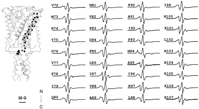
Figure 5.
X-band EPR spectra of spin-labeled mutants from the second transmembrane domain TM2 of Eco-MscL reconstituted into asolectin liposomes. The spectra were obtained using a loop-gap resonator with the microwave power of 2 mW and field modulation of 3 G. The spectra are arranged sequentially from the NH2 terminus to the COOH terminus of the MscL monomer as pointed by the arrow. The MscL subunit with mutated residues (black spheres) is superimposed onto the 3-D MscL structure (left). The scale bar corresponds to 30 G.

