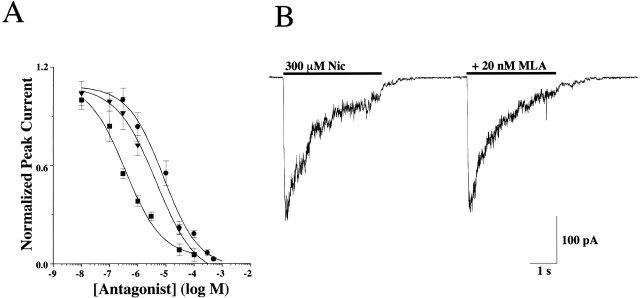
Figure 2.
Antagonist sensitivity of the AChR currents activated in IMR-32 cells. (A) The concentration response relationship is shown for d-tubocurare (d-TC; ▪), mecamylamine (Mec; ▾), and hexamethonium (C-6; •) on IMR-32 cells with IC50 values of 0.4 ± 0.2, 3.2 ± 0.6, and 8.5 ± 3.0 μM, respectively. All drugs were coapplied with 100 μM ACh at a holding potential of −60 mV. The peak amplitudes of the resulting currents were then plotted against concentration of the antagonist after normalizing to the peak amplitude of the current recorded in the absence of any antagonist. All three drugs completely blocked the currents at higher concentrations. (B) Representative currents are shown for testing the sensitivity of IMR-32 AChR currents to the reversible α7 selective antagonist MLA (20 nM). No inhibition was observed, which was consistent with the idea that the macroscopic current recorded in the cells reflects predominately the activity of α3 AChRs.

