
Figure 1.
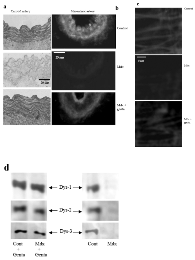
Immunolocalization of dystrophin in carotid arteries (revealed by peroxidase, a) and in mesenteric resistance arteries (Texas-red immunofluorescence, b) was performed using Dys2 anti-dystrophin antibodies. Arteries were isolated from control, mdx mice or mdx mice treated for two weeks with gentamicin. In mesenteric resistance arteries dystrophin (Dys2) was localized to the plasma membrane of endothelial cells, using immunofluorescence and confocal microscopy. This was performed in arteries perfused under a pressure of 75 mmHg and a flow of 50 μl/min (c). (n=6 mice per group). Due to the movement generated by the perfusion of the arteries in physiological conditions, the scanning of the vascular wall was performed at a speed of 10 images/sec.
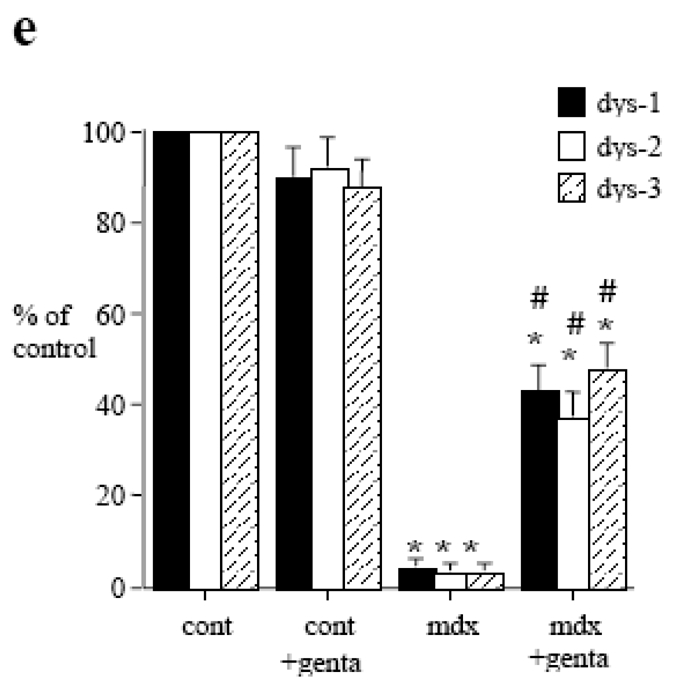
The expression of dystrophin was determined in carotid arteries, using western-blot analysis. Dys1, Dys2 and Dys3 antibodies were directed against C-terminus, N-terminus and mid-rod domains of dystrophin, respectively (d and e). (n=8 mice per group).
*P <0.001; two-factor ANOVA, versus control.
#P <0.001; two-factor ANOVA, non-treated versus gentamicin-treated mdx mice.