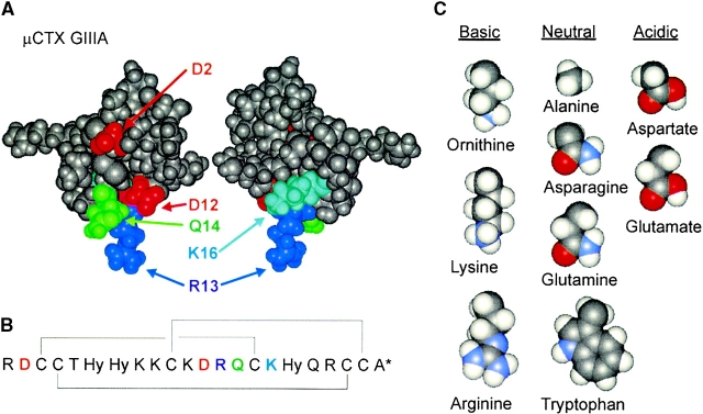
Figure 1.
Structure of μCTX. (A) Aqueous solution tertiary structure of μCTX at pH 2. The two views differ by a rotation of ∼180° around an axis defined by the R13 side chain, which is thought to enter the channel roughly parallel to the pore axis. Although the tertiary structure of μCTX is held rigidly by disulfide bonds, all charged residues point outwards from a discoidal structure of ∼20-Å diameter and are likely flexible in solution. In this study, amino acid substitutions were made at positions 2, 12, 13, 14, and 16, as labeled. Coordinates from the Protein Data Bank (structure 1TCG; Lancelin et al. 1991). (B) Primary sequence of μCTX. μCTX is a 22–amino acid peptide toxin found naturally in the venom of the Conus geographus cone snail (Gray et al. 1988). The toxin contains seven basic (+) and two acidic (−) residues. With the amidated COOH-terminal (asterisk), nominal net charge of the toxin at neutral pH is +6. The structure is held rigid by three disulfide bonds (connecting brackets) between paired cysteine residues (Price-Carter et al. 1998; Kaerner and Rabenstein 1999) and contains three modified amino acids, all hydroxyprolines (Hy; Gray et al. 1988). (C) Amino acid substitutions into position-13. Nominal side chain charge at pH 7 is indicated. Side chains are shown in space-filling representation, starting at Cα, without their backbone atoms.

