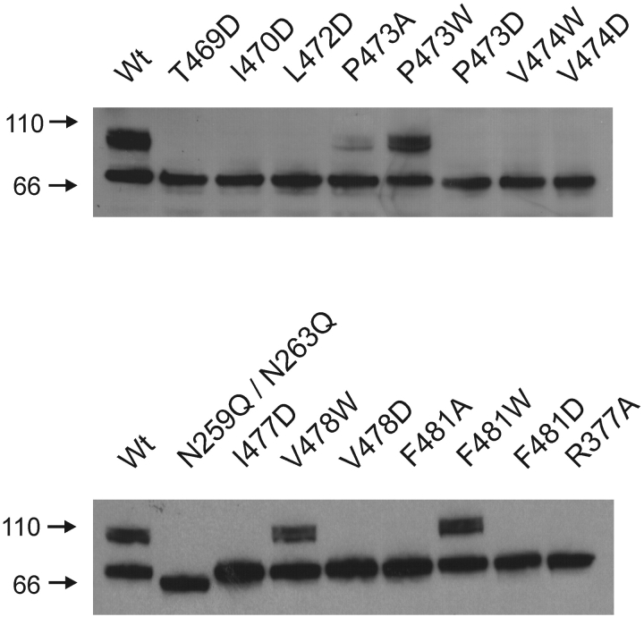
Figure 6.
Mutant Shaker Kv channels with defects in maturation. Western blots from SDS poylacrylamide gels of c-myc–tagged Shaker protein obtained from crude oocyte membrane preparations. Each lane contains between 4 to 10 oocyte equivalents of Shaker protein. Wild-type protein has two dominant forms, a core-glycosylated species (band ∼70 kD) and a more heavily glycosylated mature form (broad band at ∼100 kD). The N259Q/N263Q double mutant shows only a single band at ∼65 kD and marks the position of the unglycosylated protein. Numbers to the left are molecular weight markers in kD. See materials and methods for details.

