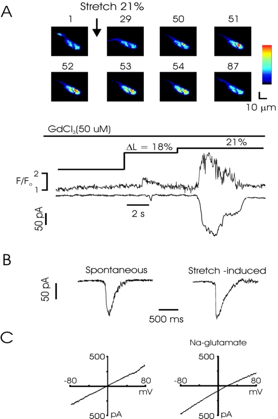
Figure 5.

Stretch-induced Ca2+ release activates STICs and macroscopic Ca2+-activated chloride currents. (A) Combined confocal and patch-clamp recording from a rabbit myocyte shows that SICR activates inward currents temporally and quantitatively linked to the Ca2+ release. The experiment was performed in the presence of Gd3+ (50 μM), which blocks stretch-activated cation currents. The numbers above each image indicate the relative timing of acquisition, beginning just before the stretch to 21%. Images were obtained every 44 ms. Pixel size = 1 μM. (B) Transient inward currents associated with calcium-activated chloride channel gating. Current at left occurred spontaneously, whereas current at right was evoked following cell stretch. Note roughly similar amplitude and kinetics. (C) I-V curve obtained from a ramp from −80 to 80 mV from a holding potential of −60 mV during stretch. Leak currents from ramp obtained before stretch have been subtracted. In physiological salt solution (left), stretch induces a linear current with a reversal potential of ∼0, the equilibrium potential for Cl−. Partial replacement of chloride with the poorly permeant anion glutamate shifts the reversal potential to ∼18 mV, close to the theoretical Cl− reversal potential of 24 mV (right).
