
Figure 8.
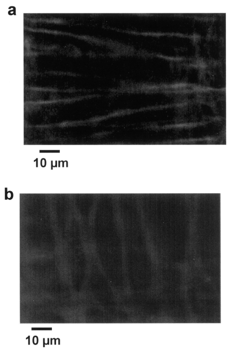
Immunolocalization of dystrophin (Texas-Red staining) in perfused resistance arteries mounted in an arteriograph (pressure = 75 mmHg and flow = 30 μl/min) shows the in situ localization of dystrophin in smooth muscle (a) and endothelial cells (b). Confocal scanning was performed at a high speed (30 images per second) due to the movements of the vessel wall during perfusion under pressure.