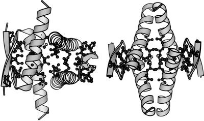
Figure 1.
Ribbon model of the human p53tet structure. Two different orientations are shown on the left and right sides. The 1sak coordinates (28) for the minimum tet domain (p53 residues 326–356), obtained from the Protein Data Bank, and the program molscript (43) were used. The residues whose side chains were found critical for stability form most of the hydrophobic core and have been depicted as ball-and-stick models.

