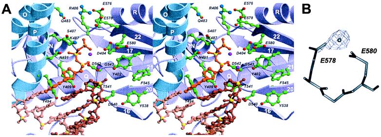
Figure 4.
Polymerase active site. (A) Stereoribbon representation (using color code as in Fig. 2) with modeled DNA. Active-site residues are shown as ball-and-stick representations with carbon (green), nitrogen (blue), and oxygen (red) atoms. The DNA template (light brown), primer (light brown), and dNTP (orange) complex has been taken from the coordinates of T7 replication complex (15) by superimposing D404, D542, and adjacent residues with corresponding residues in T7 pol (D475 and D654). Phosphorus atoms are yellow. The two metals of the T7 replication complex are shown as magenta spheres. (B) Experimentally observed metal-binding site for Mn2+ (Fo − Fc density contoured at 5σ) and Zn2+ in the “low salt” crystal form. The carboxylates E578 and E580 are conserved in type B polymerases (Fig. 3).

