
Figure 1.
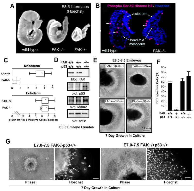
FAK-/- embryo mesoderm cell proliferation block is p53-dependent. (A) Hoechst DNA staining of wild-type (FAK+/+), FAK+/-, and FAK embryos at E8.5. (B) Mesoderm region of FAK-/- embryos lack mitotic cells. Saggital head-fold sections of E8.0-8.5 FAK+/+ and FAK-/- littermate embryos stained with phosphoserine-10 Histone H3 (red, arrowheads) and Hoechst (blue). (C) Quantitation of phosphoserine-10 Histone H3 staining. Number of mitotic cells (per headfold region section) in the ectoderm and mesoderm of FAK-/- and FAK+/+ littermates plotted as Box-and-whisker diagrams: dot (mean), box (25 to 75 percentile), and bars (minimum and maximum value). (D) Immunoblotting of embryo lysates shows increased p53 and Mdm2 protein expression from E.8.5 FAK-/- compared to FAK+/+ or FAK+/- littermates. (E) p53 prevents FAK-/- embryo proliferation ex vivo. The indicated E8.0-8.5 embryos were dissected and cultured in Matrigel for 7 days. Phase contrast images of Matrigel-embedded embryos (dark) and surrounding cells growing out from the embryo mass. (F) Rescue of FAK-/- cell proliferation defects by p53 deletion. Anti-BrdU staining (green) and Hoechst (blue) labeling of dissociated cells from embryo explants cells at 40X. Percentage of BrdU-positive cells for the indicated genotype. Data are mean +/- SEM from 3 independent experiments. (G) E7.0-7.5 time of embryo extraction does not alter FAK-/-p53+/+ proliferation defects ex vivo. Phase images of embryos in Matrigel culture for 7 days (scale bar is 100 μm). Hoechst staining shows cells with multiple (arrowheads) and fragmented (arrows) nuclei in FAK-/-p53+/+ but not FAK+/+p53+/+ cultures.