
Figure 2.
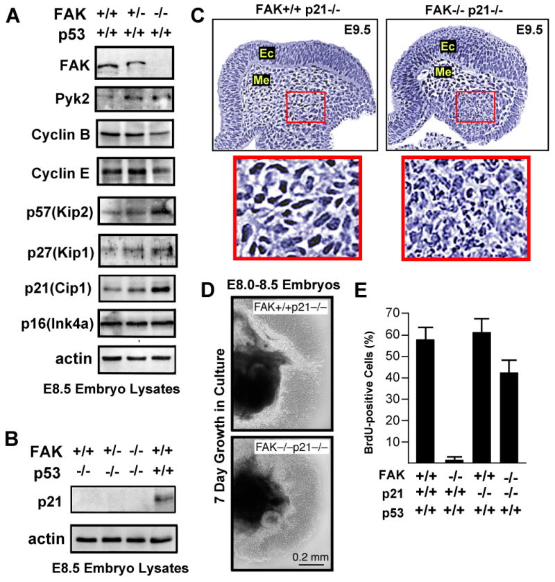
p53-mediated proliferation block of FAK-/- cells is p21-dependent. (A) Increased expression of p53, Pyk2, or cyclin-dependent kinase inhibitors and decreased expression of cyclins in FAK-/- compared to FAK+/+ littermates as determined by immunoblotting of E.8.5 embryo protein lysates. (B) Lack of detectable p21 expression in lysates from FAK-/- E8.5 embryos on a p53-/- background. (C) FAK-/-p21-/- embryos exhibit lethality at E9.5. H&E staining of saggital headfold sections. Ec=ectoderm and Me=mesoderm. Inset, fragmented nuclei observed in mesoderm region of FAK-/-p21-/- but not FAK+/+p21-/- embryos. (D) E8.0-8.5 FAK-/-p21-/- embryo cells proliferate equally to FAK+/+p21-/- cells in 7 day Matrigel culture ex vivo. Phase contrast images of embryo mass (dark) and surrounding cells. (E) p21 inactivation promotes FAK-/- embryo cell proliferation as determined by the percentage of BrdU-positive cells counted for the indicated genotype. Data are mean +/- SEM from 3 independent experiments.