
Figure 6.
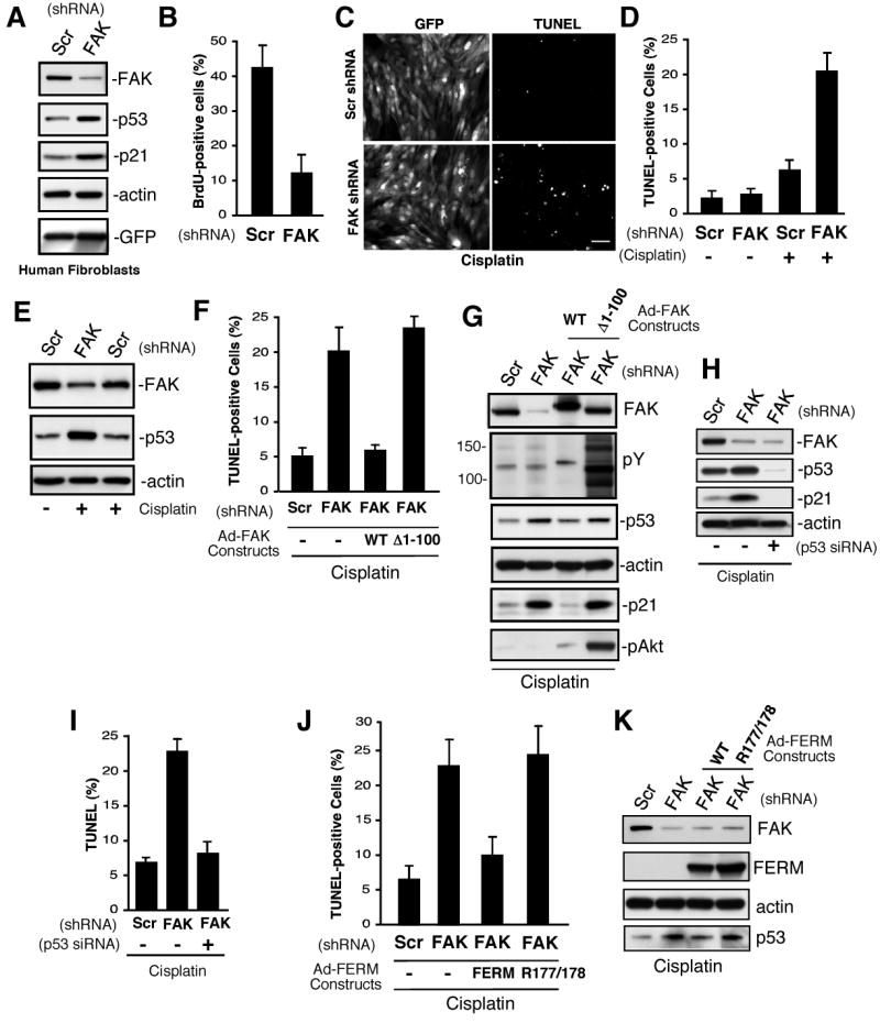
FAK controls human diploid fibroblast proliferation and p53-dependent apoptosis. (A) Lysates from scrambled (Scr) and FAK shRNA infected cells after 72 h were analyzed by anti-FAK, p53, p21, actin, and GFP blotting. (B) FAK shRNA inhibits human fibroblast proliferation. Cells were infected with the indicated lentivirus for 72 h, BrdU was added for 16h in growth media, and cells stained with anti-BrdU antibody. Mean values +/-SD are percent of total GFP-positive cells. (C, D) FAK shRNA sensitizes human fibroblasts to cisplatin-stimulated apoptosis. Cells were infected with the indicated lentivirus for 48 h, cisplatin (20 μg/ml) was added for 48 h, cells were fixed, and then analyzed by TUNEL staining. (C) Representative images of GFP-expressing and TUNEL-stained fibroblasts. Scale bar is 200 μm. (D) Mean values +/- SD for cisplatin-stimulated apoptosis were obtained by counting three TUNEL-stained 10X fields of cells from two coverslips. Only GFP-positive cells were counted and the data represents two independent experiments. (E) Elevated p53 levels in cisplatin-treated FAK shRNA-expressing fibroblasts as treated as in panel D and analyzed by anti-FAK, p53, and actin blotting. (F, G) FERM domain integrity is required for rescue of cisplatin-stimulated apoptosis. Cells were infected with Scr or FAK shRNA lentivirus (48 h), transducted with Ad-FAK or Ad-FAK (Δ1-100), and after 24h, cisplatin (20 μg/ml, 48 h) was added prior to analysis by TUNEL staining. (F) Mean values +/- SD for cisplatin-stimulated apoptosis were obtained as described for panel D. (G) FAK but not Δ1-100 FAK reverses cisplatin-stimulated increases in p53 and p21 expression as determined by blotting. Δ1-100 FAK activates Akt as determined by phospho-specific blotting. (H, I) FAK shRNA-enhanced cisplatin-stimulated apoptosis is p53 dependent. Fibroblasts were transfected with p53 siRNA, transduced with Scr or FAK shRNA lentivirus, and treated with cisplatin (20 μg/ml, 48 h). (H) Blotting for FAK, p53, and p21 blotting show changes in protein expression with actin blotting as control. (I) Mean values +/- SD for cisplatin-stimulated apoptosis were obtained as for panel d. (J, K) FAK FERM domain rescue of cisplatin-stimulated apoptosis. Cells were transduced with Scr or FAK shRNA lentivirus (48 h), infected with Ad-Myc-FERM WT or Ad-Myc-FERM R177/R178 (24 h), and then treated with cisplatin (20 μg/ml, 48h). (J) Cells were analyzed fro TUNEL staining as in panel D. (K) Blotted for FAK, Myc tag (FERM), actin, and p53 shows that FERM mutation blocks p53 regulation.