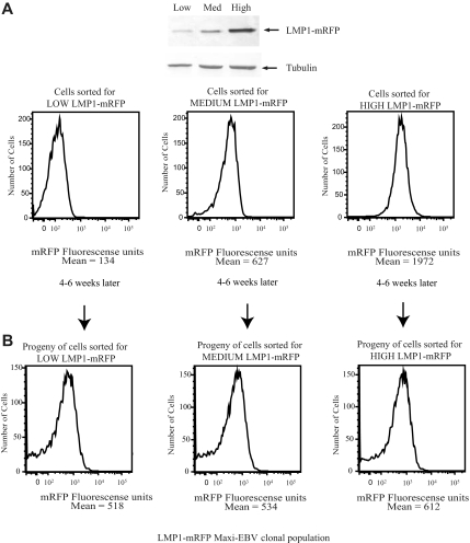
Figure 7.
Cells that express high levels of LMP1 regenerate a daughter population with a broad distribution of levels of expression of LMP1. (A) EBV-positive strains expressing LMP1-mRFP were sorted for cells expressing low, medium, or high levels of mRFP and analyzed by FACS analysis and Western blotting using anti-LMP1 and antitubulin antibodies. (B) Cells sorted for low, medium, or high levels of mRFP were plated in 96-well plates. The clones were allowed to grow for 4 to 6 weeks. Proliferating cell clones were expanded and analyzed on FACS Vantage. Data were analyzed with FlowJo software. The broad distribution of LMP1 observed in EBV-positive strain cells expressing LMP1-mRFP was confirmed to be comparable with that observed in 721 cells by fixing the cells and staining with anti-LMP1 antibodies (data not shown).

