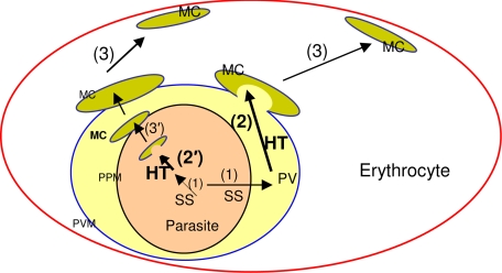
Figure 7.
Schematic for HT-mediated cleft targeting events in erythrocyte infected with the malaria parasite P falciparum. In secretory proteins, a cleavable N-terminal ER-type signal sequence (SS) delivers proteins to the PV (step 1). The HT motif enables protein accumulation in the Maurer's clefts (MC, step 2). Clefts can either bud from the PVM (also step 2) packed with proteins exported to the red cell and function as protein reservoirs underneath the erythrocyte membrane (step 3). The HT motif may also move protein from the lumen of the PVM to lumen of clefts either within the parasite (step 2′) or at the proximity to the parasite plasma membrane (PPM, step 3′). Both steps 2 and 2′ implicate recognition of the HT motif by a putative receptor located at the Maurer's clefts (not shown).

