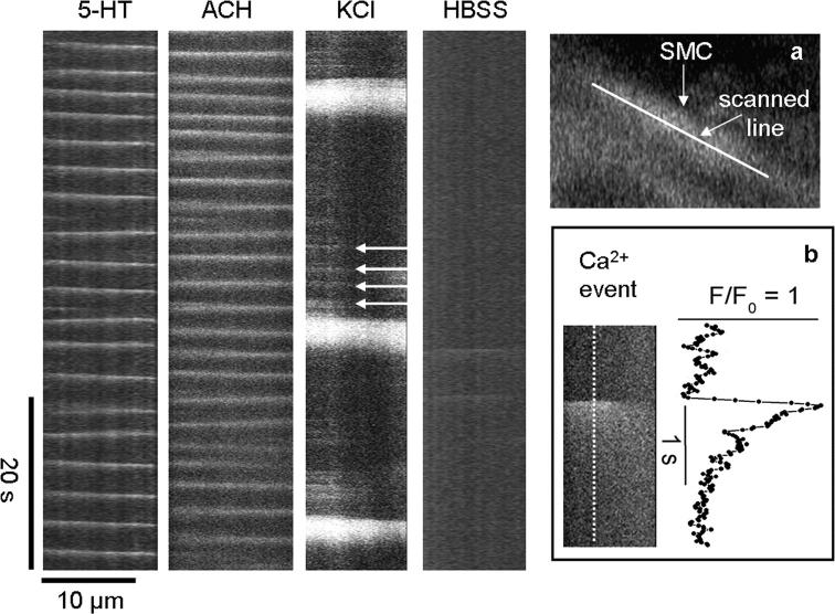
Figure 13.
Ca2+ waves and elemental Ca2+ events in airway SMCs during the stimulation with 5-HT, ACH, and KCl. Line scans from the longitudinal axes of single airway SMCs (inset, a) from high speed recordings (60 fps) of changes in Ca2+ during continuous perfusion with 1 μM 5-HT, 1 μM ACH, and 50 mM KCl and after removal of KCl by washing with HBSS, as indicated. The slopes of the white lines indicate the velocity (μm/s) and direction of the Ca2+ waves in the SMCs. KCl induce small Ca2+ events (arrows) preceding the Ca2+ wave. (Inset, b) An expanded view of a single Ca2+ event (image and trace) induced by KCl. Representative traces of at least four experiments from different slices of two mice.

