
Figure 8.
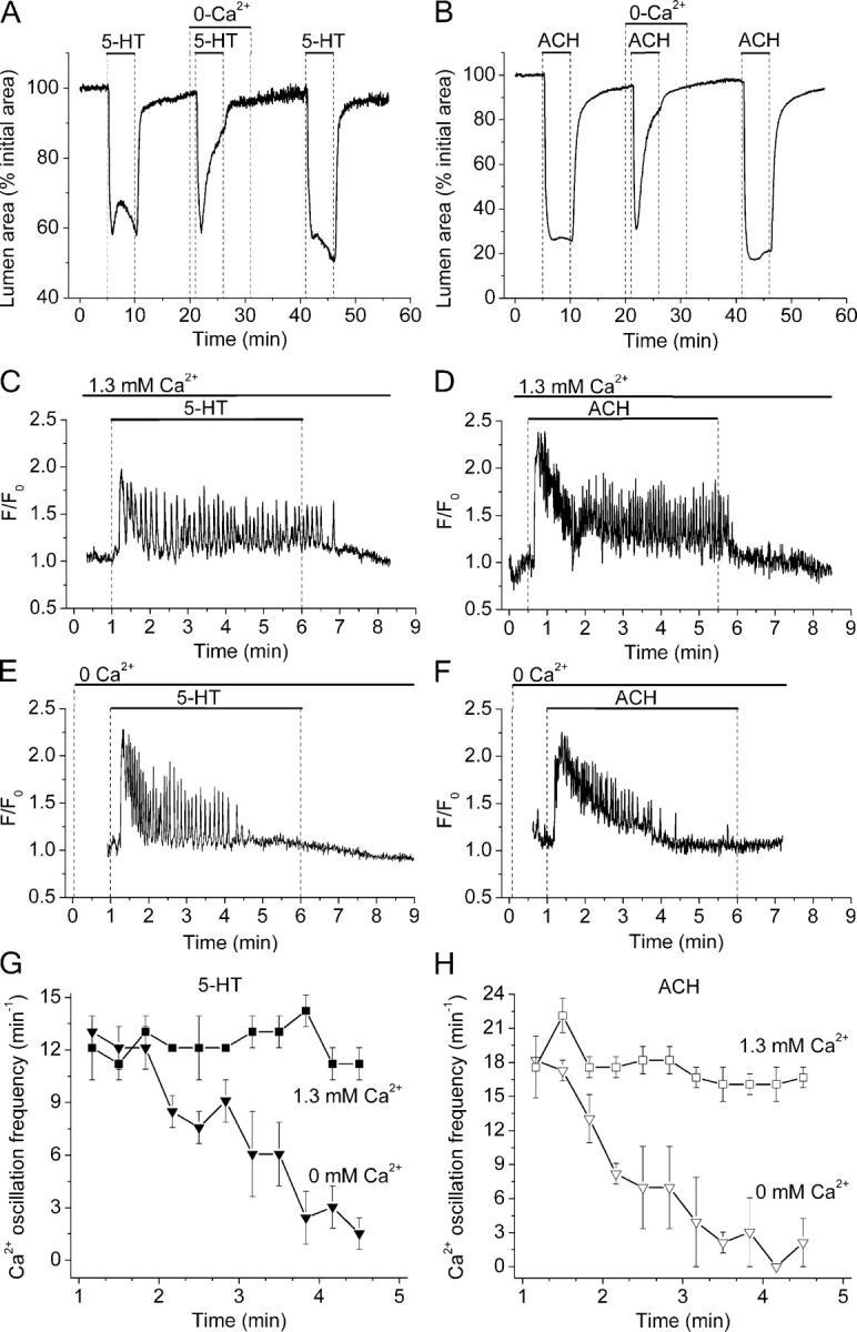
Role of extracellular Ca2+ during the contraction and the Ca2+ signaling of airway SMCs induced by 5-HT and ACH. (A and B) Contractile responses of airways in different lung slices sequentially exposed (top bars) to 1 μM 5-HT and 1 μM ACH in the presence or absence of extracellular Ca2+. Representative traces of five airways from three mice. (C–H) The changes in [Ca2+]i in small ROIs of single SMCs were determined in lung slices stimulated with (C, E, and G) 1 μM 5-HT or (D, F, and H) 1 μM ACH in the (C and D) presence or (E and F) absence of extracellular Ca2+ as indicated by top bars. In the absence or presence of extracellular Ca2+, both agonists induced an increase in [Ca2+]i followed by Ca2+ oscillations. Although Ca2+ oscillations persisted in the presence of extracellular Ca2+, they progressively reduce their frequency and stopped in the absence of extracellular Ca2+. (G and H) The frequency of agonist-induced Ca2+ oscillations, calculated every 20 s, in the presence (squares) or absence of Ca2+ (triangles) with respect to time. Symbols and bars are the mean ± SEM of five to eight SMCs in five paired experiments from different lung slices from three mice.