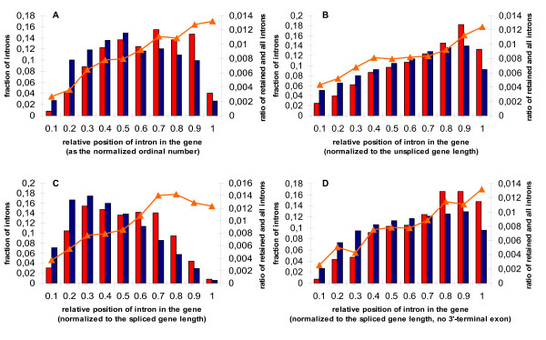
Figure 5.
Histograms of the relative intron positions. A: the relative (ordinal) intron number; B: unspliced genes; C: spliced genes; D: spliced genes with the last exon removed (see the text for the detailed explanation). Left axis: the fraction of introns in each position bin is given for retained (red) and constitutive (blue) introns separately. Points 0 and 1 on the horizontal axis correspond to the 5'- and 3'-ends of the gene, respectively. Right vertical axis and the orange triangle curve: the fraction of retained introns among all introns in the bin.

