
Figure 1.
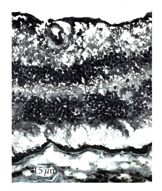
Microphotograph showing normal human retina stained for PPARγ in the ganglion cell, inner nuclear layer, outer nuclear layer, and RPE (avidin-biotin-complex immunoperoxidase).
Official websites use .gov
A
.gov website belongs to an official
government organization in the United States.
Secure .gov websites use HTTPS
A lock (
) or https:// means you've safely
connected to the .gov website. Share sensitive
information only on official, secure websites.
Microphotograph showing normal human retina stained for PPARγ in the ganglion cell, inner nuclear layer, outer nuclear layer, and RPE (avidin-biotin-complex immunoperoxidase).