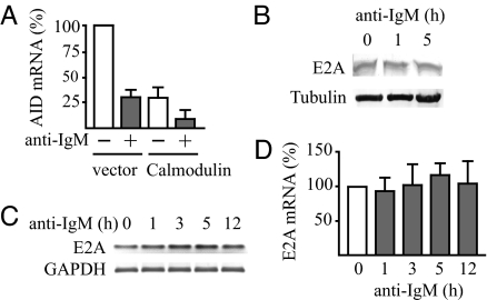
Fig. 2.
Inhibition of AID gene expression by calmodulin and constant E2A mRNA and protein expression after engagement of the BCR with anti-IgM. (A) Down-regulation of the AID mRNA level by calmodulin overexpression with and without inhibition of the gene by BCR engagement. DG75 cells were transfected with 2 μg of pcDNAI/amp (vector) or pcDNAI/amp mCaM (Calmodulin) plasmid. Eight hours later, live cells were separated by using Lymphoprep (Axis-Shield), and, 12 h later, anti-IgM was added to one-half of the cells followed by continued culture for 3 h. Expression levels of AID mRNA were determined by quantitative RT-PCR and normalized by using GAPDH as in Fig. 1. The level before addition of anti-IgM in cells transfected with the pcDNAI/amp vector was set at 100%. Results are mean ± SD (n ≥ 3). (B) Levels of E2A protein in anti-IgM-treated DG75 cells determined by Western blot analysis. Levels of α-tubulin were used as loading controls. (C) E2A mRNA levels of anti-IgM-treated DG75 cells determined by PCR and agarose gel electrophoresis of synthesized cDNA strands as in Fig. 1. (D) E2A mRNA levels of the anti-IgM-treated DG75 cells determined by quantitative RT-PCR as in Fig. 1. Results are mean ± SD (n = 3).

