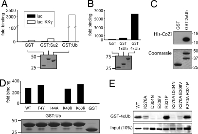
Fig. 3.
IKKγ binds Ub via its CoZi domain. (A and B) Purified GST fusion proteins coupled to beads were incubated with lysates of 293 cells expressing luciferase IKKγ. The ratio between luciferase activity bound to beads and present in lysates is shown. GST fusion proteins were visualized by Coomassie blue staining. (C) Purified GST fusion proteins coupled to beads were incubated with lysate from bacteria expressing His-tagged CoZi. (Upper) Eluates from beads were blotted with anti-His antibody. (Lower) GST fusion proteins were visualized by Coomassie blue staining. (D) Purified GST fusion proteins coupled to beads were incubated with lysates of 293 cells expressing a luciferase IKKγ. The ratio between luciferase activity bound to beads and present in lysates is shown. GST fusion proteins were visualized by Coomassie blue staining. (E) The 293 cells were transfected with the indicated AU1-tagged IKKγ constructs. Lysates were incubated with purified GST-tetra-Ub (GST-4xUb) bound to beads. Lysates and eluates were blotted for AU1-IKKγ.

