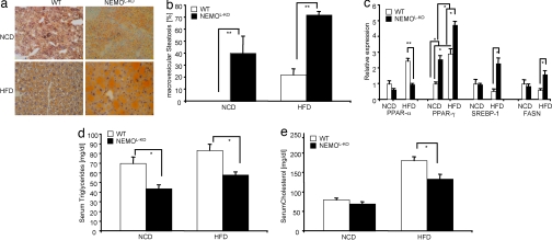
Fig. 2.
Macrovesicular steatosis in livers of NEMOL-KO mice. (a) Representative Sudan staining from liver sections of control and NEMOL-KO mice on NCD and HFD at 5 months of age. Sections were counterstained with hematoxylin. (b) Percentage of macrovesicular lipid accumulations in lipid containing hepatocytes. (c) Relative expression of the indicated genes from the different genotypes was performed by real-time PCR by using TaqMan Assay (n = 8 per genotype). Values are mean ± SEM. *, P ≤ 0.05; **, P ≤ 0.001 vs. control. (d) Triglyceride and (e) cholesterol levels from sera of control and NEMOL-KO mice on NCD and HFD at week 16 were determined by a diagnostic laboratory using standard techniques (n = 16 per genotype).

